The heart is a fist-sized organ located in the front of the chest. Made up of muscle and tissue, it is the main organ of your circulatory system, and its primary function is to move blood throughout the body. Blood brings oxygen and essential nutrients to all parts of the body, including the cells and organs, so they can keep working and stay healthy. Heart conditions are among the most common types of disorders and can affect your quality of life and wellbeing.
An MRI can help in the assessment and treatment of several heart conditions by creating detailed images of the inside of your body in two or three dimensions. It looks specifically at the heart, as well as the surrounding blood vessels and how your blood moves. The high-resolution pictures help your healthcare provider figure out what is wrong and make an accurate diagnosis of various conditions related to the heart.
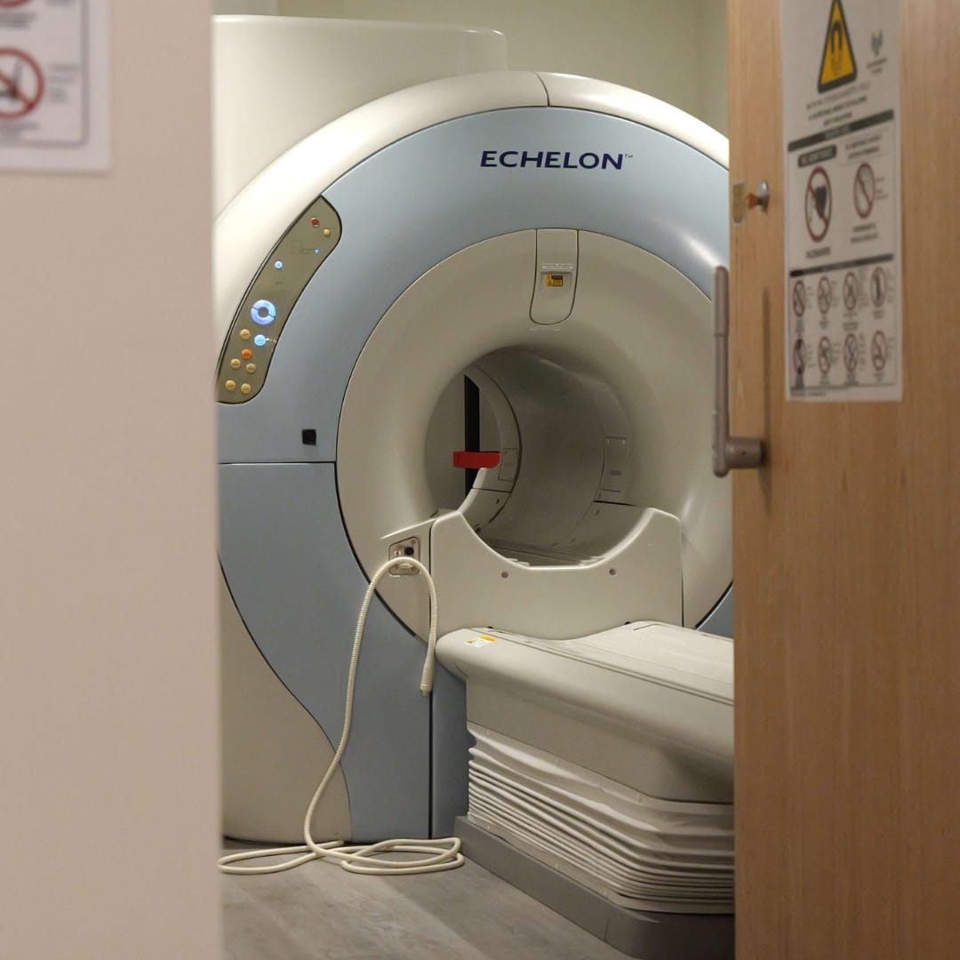
 Cardiovascular MRI is an advanced, non-invasive imaging technique that uses a strong magnetic field, radio waves, and a computer to create very fine, detailed images of the structures within and around the heart, including chambers, valves, and muscles. It allows the doctor to evaluate the size and shape of your heart as it shows the thickness of your heart muscle and the function of the heart valves.
Doctors use cardiac MRI to detect or monitor cardiac disease. They also use it to evaluate the heart’s anatomy and function in patients with heart diseases present at birth and heart diseases that develop after birth. By identifying any potential issues early on, an MRI scan helps physicians develop a personalized treatment plan to improve heart health and prevent further complications.
An MRI may be used instead of a CT scan, particularly when organs or soft tissues are being studied. A cardiovascular MRI may provide the best images of the heart for certain conditions as it does not use radiation like X-rays or CT scans.
An MRI of the heart is usually requested if you have a more complex or advanced heart condition, often after initial first-line testing, such as transthoracic echocardiography. MRI images show the parts of your heart and any damage to specific areas. It also indicates how well your heart’s chambers and valves are working, how the blood is moving, and detects areas of damaged or diseased tissue.
Your doctor may order a cardiac MRI to assess signs and symptoms that suggest:
Your physician may also ask for a cardiovascular MRI to plan cardiac procedures such as coronary artery bypass surgery, pacemaker or defibrillator implantation, and catheter ablation for atrial fibrillation.
Your doctor may refer you for an MRI scan if you are experiencing some of the following symptoms:
These symptoms can be a sign of heart disease or heart failure, which is a condition when the heart is unable to pump enough blood to meet the body’s needs. An MRI enables the doctor to further investigate your heart health, determine heart muscle damage, heart valve problems, and inflammation, and make an accurate diagnosis regarding your condition.
A cardiovascular MRI is also used to track the progress of heart conditions over time and assess the effectiveness of treatment.
There is no special preparation for a cardiac MRI, but you should follow the guidelines provided by your doctor.
Inform the technologist if you have any devices or metal in your body from your previous surgery or procedure in your body, including:
You can undergo an MRI with implanted cardiac pacemakers and defibrillators, but it requires special consideration based on the type of device you have and the MRI equipment. It is essential to provide complete information about the implanted device to the technologist when scheduling a scan to prevent delays and other complications.
An MRI is a painless, simple process, but lying still for some time may cause discomfort, especially if you are not feeling well or have been through some invasive surgery. Inform the technologist about your condition, and they will use all possible measures to ensure your comfort and complete the scan as quickly as possible.
There is no post-MRI care. However, you must be careful and move slowly when getting up from the scanner table to avoid dizziness or lightheadedness from lying flat for some time.
If you were given an anti-anxiety pill or sedated for the scan, you need to rest until its effects have worn off. You will need someone to drive you home afterward. You will also be monitored for reactions, such as itching, swelling, rash, or difficulty breathing if contrast dye was used for your scan. They are rare and usually mild and can be managed with medication.
You can go back to your routine diet, medication, and activities, unless you have other instructions from your doctor, depending on your heart condition. If you notice any pain, redness, or swelling at the IV site after you have left the facility, call your doctor.
A radiologist will review and analyze the images, prepare a report, and send it to your referring physician who will discuss the test results with you. You can also request a copy of images on a CD ROM for your record.
If a follow-up examination and treatment is required, your doctor will tell you about it. It may be a significant step for further evaluating a potential issue, planning the next step in your care, or monitoring changes over time.
Cardiac MRI scans are safe, risk-free procedures, but there may be other risks depending on your specific medical condition.
Cardiac MRI uses special techniques to create detailed pictures of the heart to specifically look at its structure and function, as well as the surrounding blood vessels to determine your heart condition. Learning what happens during the scan and how to prepare for it makes the process less daunting and helps you go through it smoothly.
The cost of a cardiac MRI in Midtown Manhattan depends on factors such as the use of contrast and your insurance coverage. Typically, the price ranges from $450 to $2,500, with our cost $450. Manhattan MRI accepts most insurance plans, which can substantially reduce or fully cover the cost of your cardiac MRI.
Has your doctor ordered a cardiovascular MRI to assess your heart condition? If yes, call Manhattan MRI today and schedule a scan so your physician can get a comprehensive, accurate look at your heart without any invasive test or surgery. Our specialists help you understand what this scanning process includes and how to prepare for it to ensure the best possible outcomes that restore your quality of life.