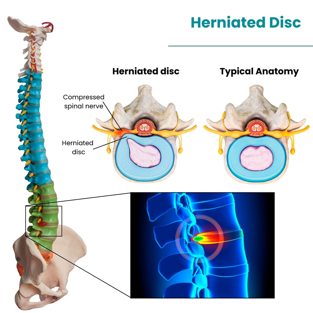
Herniated discs are a common cause of low back pain and usually result from accidents, sports-related activities, or spinal wear and tear due to aging. Disc herniation most often occurs in the lower area of the spine, also known as the lumbar spine. Lumbar disc herniation is one of the most common causes of lower back pain and sciatica.
An MRI of the spine can help to diagnose and monitor herniated spines. It can be used in a variety of ways, including to locate the herniated disc and determine the level of nerve compression.
 A herniated disc occurs when one of the discs between the vertebrae in the spine gets damaged. These discs act as shock absorbers and keep the vertebrae from rubbing against each other. The protrusion or herniation of the disc can lead to various symptoms depending on the area of the spine that is affected. Several terms are used to describe a herniated disc, including bulging disc and pinched nerve. The symptoms are often similar, often making diagnosis a challenge.
While many cases of disc herniation tend to improve with conservative measures within a few weeks, MRI is recommended for people with severe pain, neurological symptoms suggestive of spinal cord compression, or chronic pain that has lasted for more than six to eight weeks.
An MRI scan is an imaging test that uses radio waves to generate high-quality pictures of the body area being scanned. It has proved to be the best imaging test for evaluating a herniated disc, with a diagnostic accuracy of 97%. An MRI of the spine can visualize spinal bones and the surrounding soft tissues, including ligaments, muscles, blood vessels, and nerves.
MRIs allow your healthcare provider to diagnose a herniated or bulging disc. It is a valuable tool that helps physicians check whether your pain is caused by one slipped disc or if multiple degenerative discs are limiting your range of motion. They can better visualize the affected area for pre-operative planning when surgical intervention is required.
It is one of the safest and most effective tests as it does not pose any radiation risk, like X-rays and CT scans.
Chronic back pain that does not get better with rest and medications and radiates down to your arms or legs indicates a serious concern. Your doctor may recommend an MRI to view the spine and determine if there is a herniated disc, and in some cases, find out what caused it.
An MRI offers visual confirmation of the bulging disc, taking detailed images of the parts of the body that makes up the spinal column and the spinal discs between the vertebrae. With the right imaging, they can see if any discs are herniated and the severity of their movement into your nerve canal.
An MRI is a crucial part of a herniated disc diagnosis. It does not require any special preparation, but it is essential to follow the guidelines and instructions provided by your doctor to ensure accurate results.
Inform your doctor about any mental implants you have from previous surgery or any devices in your body, such as:
It may not be safe for you to have an MRI scan as the magnetic field the scanner produces is extremely powerful and may interact with any metal in or on your body.
An MRI is no more painful than getting your picture taken, but staying still for some time can cause some discomfort, particularly in case of a recent injury or an invasive procedure such as surgery. The technologist will strive to complete the process as quickly as possible to minimize pain.
There is no special post-MRI recovery or care unless you took anti-anxiety medication to stay calm during the scan. Once the procedure is complete, you will be allowed to get up, change into your clothes, and go home. However, it is better to get up slowly from the table and move cautiously to avoid dizziness or lightheadedness from lying down for some time, which can cause further pain in your back.
Let the technologist know if you are feeling side effects or reactions from the contrast dye used for the scan. You can return to your normal diet, activities, and medication unless you have been provided additional instructions by your doctor regarding your specific condition.
Once the procedure is complete, a radiologist will prepare the report and share a copy with your doctor who recommended the MRI. Your doctor will discuss the results with you and explain what they mean. You can also access your report online or ask for the images to be transferred onto a CD ROM for your record.
Depending on the results, your doctor might recommend more tests or discuss the treatment plan if they have a confirmed diagnosis.
Because ionizing radiation is not used, there is no risk of exposure to radiation during an MRI procedure. However, there may be other risks based on your health or medical condition.
Herniated disc MRI is a non-invasive, simple diagnostic procedure that does not take long. It helps your doctor identify the problem timely, increasing the likelihood of successful treatment. Knowing what to expect before, after, and during the scan not only makes the process less intimidating but also gives you a chance to feel more confident about what you will be going through.
Does your doctor suspect your back pain may be resulting from a herniated disc – Call Manhattan MRI today to schedule a scan. A herniated disc MRI can help your doctor pinpoint the cause of pain and rule out another diagnoses to alleviate your pain and discomfort. Our specialists help you every step of the way, address your concerns, and ensure the highest quality scanning services that result in quick and effective treatment.