The prostate gland is a small, soft structure about the size and shape of a walnut. A part of the male reproductive system, it lies deep in the pelvis between the bladder and the penis, and in front of the rectum. Its function is to liquefy semen produced from the male sexual organs to fertilize the female egg.
An MRI is a powerful, effective tool for screening and visualizing soft tissue structures in the body. The detailed MRI images allow doctors to examine the prostate gland and surrounding tissues and diagnose medical conditions.
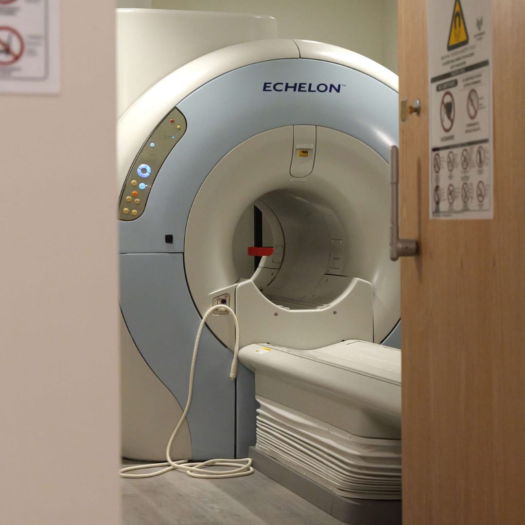
 A magnetic resonance imaging (MRI) scanner uses strong magnetic fields to create an image of the prostate and its surrounding tissues. It is the best, non-invasive method for diagnosis, local staging, and detection of prostate issues. The images can reveal tiny changes in the prostate that suggest cancer, playing a pivotal role in early detection. It may even help find lesions before symptoms arise.
MRI provides information on how water molecules and blood flow through the prostate. The prostate produces PSA, which can be measured in a blood sample. If you have prostate cancer, your PSA level may be high. However, it may be higher for another reason, such as an infection of the prostate gland or prostatitis.
MRI of the prostate is needed to evaluate other prostate issues, including infection or abscess. An MRI greatly increases accuracy and ultimately improves patient care.
Your doctor may order an MRI of the prostate gland for several reasons, including:
MRI is the most effective way to obtain detailed images of the prostate gland as compared to other radiological tests such as CT scan or ultrasound. It can tell the difference between diseased and normal tissue, which make early cancer detection possible. It also enables the physician to discern whether the cancer has extended beyond the prostate and if it is aggressive to come up with the best treatment strategy.
Not only does it evaluate the prostate and identify tumors, but an MRI also guides the urologist to target those specific areas to biopsy them correctly. The improved accuracy of tumor detection also reduces the number of biopsies needed. Following treatment, MRI imaging of the prostate can help to identify potential areas of residual disease and help in subsequent treatment planning.
There is no special preparation for the scan. However, following the instructions or guidelines provided by your doctor is essential to ensure accurate results.
Inform your doctor if you have any of the following from your previous surgeries or procedures before the MRI:
Some of these may not be compatible with the strong magnetic field of the MRI scanner and can cause you harm or suffer damage in the MRI machine. Take any documents or information about the metal implants or electronic devices inside you to assist the radiologist in deciding if your scan can be performed safely.
There is no special post-MRI care as there are no side effects to the exam. Once the procedure is complete, you can get up from the table, change back into your clothes, and go home. Some people may experience a dry mouth and mild blurring of vision as a result of the medication given for controlling bowel movement, but it does not last long.
Inform your technologist if you feel a reaction from the contrast dye used for the scan, such as nausea, itching, or hives. If you were given an anti-anxiety medication or sedative for the test, you will be monitored until you are back to normal.
You can resume your routine diet, activities, and medication unless your doctor has instructed you otherwise depending on your condition. There may be a few drops of blood after the endorectal coil is removed, especially if you underwent a recent prostate biopsy. Call your doctor if this occurs.
The images produced during the MRI scan are interpreted or read by a specialist doctor, called a radiologist. The radiologist will prepare the report and share it with your referring doctor, who will share the results with you and explain what they mean. You can also request a copy of the images on the CD ROM for future reference.
You can expect the results of your prostate MRI based on the urgency with which the result is needed. Your doctor may recommend more tests or discuss the next step with you, depending on your MRI result and your condition.
An MRI of the prostate plays a crucial role in identifying cancer of the prostate gland, especially if you have elevated or rising PSA.
You may benefit from prostate MRI if you have two or more risk factors:
If your cancer has already been detected, the MRI images can show whether it has spread outside the prostate gland or not. This can have a significant impact on your treatment and recovery.
Some people have an allergic reaction to the contrast dye that is used for highlighting certain areas of the body during the scan. You may experience rash, hives, or nausea for some time. There may be a risk of excessive sedation if you took an anti-anxiety medication to stay relaxed during the scan. You will be monitored till the effects of the medication wear off. You should have someone to drive you home afterward.
In case you suffer from kidney problems or have very poor kidney function, you will not be given a contrast as there is a small risk of nephrogenic systemic fibrosis.
If an endorectal coil was used for the scan, there is a very small risk of damage to the rectum from the balloon. If you notice any blood, call your doctor.
A prostate MRI is a non-invasive way to examine your prostate and helps your physician learn more about your medical condition. Knowing what happens and how you can prepare for it makes the process less stressful and easier to go through and you can work with your technologist to obtain the most accurate results.
Has your doctor ordered a prostate MRI for you? If yes, don’t delay it, as it can help with timely diagnosis and management of your condition. Call Manhattan MRI today and schedule an appointment to learn more about your prostate health. Our specialists understand that you may have some concerns regarding the procedure and its results, and offer comprehensive information, as well as the highest quality, patient-focused care that eases your mind.