The spine plays a fundamental role in supporting our body. It protects the spinal cord, enables movement, and connects different parts of our musculoskeletal system. Injuries, genetics, congenital conditions, as well as infections, and lifestyle habits, can result in spinal problems. Seeking medical attention for persistent back pain or spinal issues can help initiate the healing process, and reduce the risk of developing a more serious condition.
An MRI is a non-invasive and highly accurate screening method that helps doctors visualize your spine’s structures and evaluate them for any abnormalities. It uses a combination of powerful magnetic field, radiofrequency pulses, and a computer to produce detailed pictures of organs and structures within the body.
What Is an MRI of the Brain or an MRI of the Spine?
 Magnetic resonance imaging, or simply MRI of the spine and brain, helps your doctor see detailed pictures of your brain, spine, spinal cord, and surrounding tissues. It is an effective diagnostic tool that can identify inflammation, bleeding, nerve root compression, and other problems in the cervical or upper spine, thoracic or mid-spine, and lumbar or lower spine.
You may need an MRI if you have been experiencing symptoms and the doctor is working to diagnose your condition. MRI produces very clear, detailed images of the specific part of the body to help your doctor learn more about your health.
Currently, MRI is the most sensitive imaging test available for the spine, especially when organs or soft tissues are being studied. It can better detect subtle changes in the vertebral column and differentiate between normal and abnormal soft tissue.
Reasons for an MRI of the Spine or Brain Why May Your Doctor Ask for a Spine MRI
Doctors order an MRI to examine the brain and spinal cord to look for injuries or the presence of structural abnormalities or certain other conditions, including:
- Tumors
- Abscesses
- Congenital abnormalities
- Aneurysms
- Venous malformations
- Hemorrhage, or bleeding into the brain or spinal cord
- Subdural hematoma (an area of bleeding just under the dura mater, or covering of the brain)
- Degenerative diseases, multiple sclerosis, hypoxic encephalopathy (dysfunction of the brain due to a lack of oxygen), or encephalomyelitis (inflammation or infection of the brain or spinal cord)
- Hydrocephalus, or fluid in the brain
- Herniation or degeneration of discs of the spinal cord
- Surgeries on the spine, such as decompression of a pinched nerve or spinal fusion
Spine MRI can help to identify the specific location of a functional center of the brain, a part of the brain that controls functions such as speech or memory to assist in treatment. It aids in detecting other possible causes of back pain, such as compression fractures and bone swelling.
Additionally, if you are already receiving treatment for an illness or injury, your doctor might use an MRI to determine how well the treatment is working. They will get an idea of how your body is recovering and could monitor changes in the spine or brain after an operation, such as scarring or infection.
What Are the Risks of an MRI?
While the process does not have any risk to the average patient when safety guidelines are properly followed, there may be risks depending on your specific medical condition.
- As MRI does not involve radiation, there is no risk of exposure to ionizing radiation during an MRI exam. However, due to the use of strong magnets, special precautions are taken when MRI is performed on patients with certain implanted devices, such as pacemakers or cochlear implants.
- The MRI technologist will collect information regarding your implanted device, such as the make and model number to determine if it is safe for you to have an MRI. Patients with internal metal objects, including surgical clips, plates, screws, or wire mesh, might not be eligible for an MRI.
- If you feel unsettled in enclosed spaces, ask your physician to provide an anti-anxiety medication before the MRI exam. Make sure someone is with you to drive you home after the procedure.
- If you are pregnant or suspect you may be pregnant, notify your healthcare provider. There is no data to confirm that MRI is harmful to an unborn child, but MRI testing during the first trimester is discouraged.
- Your doctor may ask for a contrast dye to be used during the MRI exam to help the radiologist get a better view of internal tissues and blood vessels on the completed images. With contrast dye, there is a risk for allergic reactions such as hives, nausea, or itching. If you have sensitivity to contrast dye or iodine, inform your radiologist.
- Be sure to discuss any concerns with your doctor before the procedure if you have any special needs or health issues, particularly if you cannot lie down for 30 to 60 minutes at a stretch.
How to Prepare for an MRI?
MRI exams require little preparation. Follow your doctor’s instructions and stay calm to ensure your scan goes smoothly.
The following information helps you prepare for your spine MRI:
- Eating & drinking – You may eat, drink, and take your medications as usual for most MRI exams. However, some specialty MRI exams have certain restrictions. Your doctor will provide detailed instructions if you need to refrain from consuming any food or drink a few hours before the scan when you schedule your exam if needed.
- Anxiety medication – You will be lying in an enclosed tube-shaped machine during the MRI. It can take some time, as long as an hour, to complete the scan. If you have ever dealt with claustrophobia, you will need some anti-anxiety medication to cope with the process. Talk to your doctor ahead of time, and explain your concerns as well as your history with claustrophobia. They will prescribe a medication you can take before the procedure starts to help you stay calm during the MRI.
- Strong magnetic environment – Tell the technologist if you have devices or metal in your body as they might interfere with the magnetic field inside the MRI machine and result in distorted images and inaccurate results. It is best not to apply any deodorants, antiperspirants, perfumes, or body lotions before the examination, as they may contain metal and could end up delaying or rescheduling your scan.
Based on your medical condition, your healthcare provider may have to make specific preparations to ensure they get the most accurate results.
When you call us to make an appointment, it is extremely important to tell your doctor if you have any of the following:
- A pacemaker or artificial heart valves
- Any type of implantable pump, such as an insulin pump
- Vessel coils, filters, stents, or clips
- Body piercings
- A medication patch
- Permanent eyeliner or tattoos
- Metallic fragments anywhere in the body
- Experience of working with metal, such as a metal grinder or welder
What Happens During an MRI?
Generally, MRIs follow this process:
- You will be asked to remove all clothing, jewelry, eyeglasses, watches, hearing aids, hairpins, removable dental work, or any other object that can interfere with the procedure. Nothing can be worn during the scan. You will be given a gown to wear for the scan.
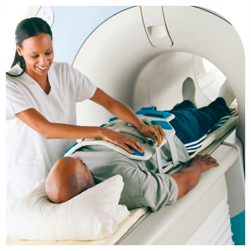
- The MRI machine is a large, tube-like structure, open on both ends. A scan table slides into a large circular opening of the scanning machine. For a spine MRI, you will be moved into the scanner head first. During the scan, you have to lie still for quality images.
- The technologist will be in another room where the scanner controls are located. He or she will be watching you through a window during the scan. Speakers inside the scanner enable the technologist to communicate with you and hear you. You will have a call button to let the technologist know if you have any problems at any time during the procedure.
- As MRI machines are noisy, you will be given earplugs or a headset to block the sounds. Some headsets also provide music for you to listen to while the scan takes place.
- During the scanning process, you will hear a clicking noise as the magnetic field is created and pulses of radio waves are sent from the scanner. It is normal and indicates the MRI machine is at work.
- You must remain very still during the examination. Any movement could cause distortion and affect the quality of the images.
- At some point during the scan, you may be asked to make small movements for better scanning, hold your breath, or not breathe for a few seconds.
- If your MRI will be done with contrast, an intravenous (IV) will be started in the arm or hand in which the contrast dye will be injected. When contrast dye is injected into the IV line, you may feel a flushing sensation or a feeling of coldness, a salty or metallic taste in the mouth, a brief headache, itching, or nausea or vomiting. These effects last for a few moments.
- If you do not feel well or experience breathing difficulties, sweating, numbness, or heart palpitations, let the technologist know immediately.
- The scan can last from 15 minutes to 60 minutes, depending on the area being scanned and number of images that are taken.
Once the scan is complete, the table will slide out of the scanner. You will be assisted off the table. If an IV line is hooked for administrating contrast, it will be removed.
The MRI procedure is not painful, but as you have to lie still for some time, it can cause some discomfort or pain, particularly if you have been through a recent injury or an invasive surgery. The technologist and other staff ensure your comfort and strive to complete the procedure as quickly as possible to minimize pain or discomfort.
What Happens After an MRI?
- There is no special care required after an MRI scan of the brain or spine. However, as you get up from the scanner table, move slowly and carefully to avoid any dizziness or lightheadedness from lying flat for the length of the procedure.
- The radiology staff will bring you back to your locker, and you can change back into your clothes.
- If you took any anxiety-relieving medication for the procedure, you may be required to rest until the effects of the medication have worn off.
- If contrast dye was used for the procedure, you may be monitored for some time for any side effects or reactions such as itching, swelling, rash, or difficulty breathing.
- If you notice any pain, redness, or swelling at the IV site after you return home following your procedure, notify your doctor as this could be a sign of infection or other type of reaction.
- You can resume your routine diet and activities, unless your doctor has advised you differently, considering your condition or specific situation.
Results and Follow-up
After your MRI scan, a radiologist will analyze the images. Once the report is ready, the radiologist will send a copy to your primary healthcare provider or the referring physician, who will share the results with you. You can also access your report online or have the images copied on a CD ROM for your record.
Once you know what to expect before, during, and after an MRI, the process becomes much less intimidating, and you feel prepared for what is to come. Not only it is painless and virtually risk-free, but also provides doctors an invaluable insight into what may be going on inside your body. It is an essential step for diagnosing an ailment and assessing how well the current treatment is progressing.
How Much Does a Spine MRI Cost in Midtown Manhattan?
The cost of an MRI of the spine in Midtown Manhattan varies depending on factors like contrast use and insurance coverage. Generally, the price ranges from $500 to $1,800, with the average cost around $1,000. Manhattan MRI accepts most insurance plans, which can significantly reduce or fully cover the cost of your spine MRI.
Have you been recommended a spine MRI by your doctor? If yes, there is no reason to wait. Call Manhattan MRI today and request an appointment with our specialists if you have any concerns or questions regarding the procedure. Our team of specialists is prepared to answer your questions, address your apprehensions, and assist you throughout the entire process most compassionately and professionally.
 Magnetic resonance imaging, or simply MRI of the spine and brain, helps your doctor see detailed pictures of your brain, spine, spinal cord, and surrounding tissues. It is an effective diagnostic tool that can identify inflammation, bleeding, nerve root compression, and other problems in the cervical or upper spine, thoracic or mid-spine, and lumbar or lower spine.