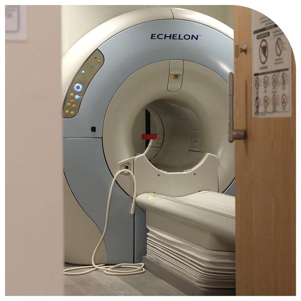
Has your doctor ordered an MRI to learn more about your pain, unusual symptoms, an underlying condition or to monitor your treatment? Contact Manhattan MRI today and discover more about MRI scans, how an MRI with contrast works, and schedule a scan with us today. Our team of specialists understands that each patient is unique with their specific health needs and recommends the type of imaging they need. With state-of-the-art imaging equipment, highly trained staff, and clear, detailed images, you can look forward to getting the most accurate diagnosis to make significant decisions regarding your health and treatment.
 An MRI scan helps in diagnosing or ruling out a disease or medical condition. It enables the physicians to determine an injury and the extent of damage it has done. It is also used for monitoring your response to treatments and keeping an eye on your recovery.
Magnetic resonance imaging technology has revolutionized the world of medical diagnostics, providing physicians with consummate insight into the human body. These days MRI scans are used for detecting, evaluating, and treating a wide range of medical conditions, including stroke, cancer, and sports injuries.
MRIs are unique to other imaging methods as they could involve using contrast agents, also known as contrast media, to enhance the visibility of internal structures.
There are two types of MRI imaging:
A contrast agent is a liquid injected into your body to distinguish between different tissues and structures in the body or to make certain tissues clearer during the imaging process. It alters the way electromagnetic radiation or ultrasound waves pass through the body and makes one structure more visible than the surrounding tissues.
Your physician will determine which type of MRI imaging is best for you and provide detailed images they need to make the best decisions regarding your health.
Read on to learn more about MRI with and without contrast, and how understanding the difference between these two can help you better prepare for your scan and ensure the most accurate imaging results.
A contrast agent also called a contrast dye, is used in an MRI to create detailed images of the inside of your body. Doctors and radiologists usually use gadolinium-based contrast agents for MRIs as they are non-toxic, extremely safe, and eliminated easily from the body.
A contrast scan helps the radiologist detect even the smallest distinctions in the tissues, such as differences between healthy and diseased tissue. Keeping this in mind, contrast is used for evaluating specific issues such as a tumor or cancer, infection, inflammation, an abscess, or an inflammatory lesion like the one caused by multiple sclerosis.
To obtain the most accurate images, a patient is first scanned without a contrast agent and then scanned again after receiving the contrast agent. This way, the radiologist can determine if the contrast has caused any unusual changes in the scan. If there is a cancerous mass or infectious abscess, it will absorb the contrast material, making it more visible against normal tissues.
Non-contrast MRIs are the ones that produce images of the body without using contrast dye. An MRI can create accurate, reliable, and detailed images even when a contrast agent is not used. It can identify a large tumor or other abnormalities when they are of a certain size.
However, not all MRI scans require a contrast agent to address the problem of concern. About 85% of MRI scans are performed without contrast. An MRI scan, also known as magnetic resonance angiography, is used to create images of the blood vessels. These scans are performed without contrast as the MRI machine can detect changes in the vascular flow, such as narrowing or blockage, without the need for contrast dye.
It is important to note that while contrast agents can offer better clarity and enhanced images in certain situations, MRIs without contrast have their own advantages.
The difference between contrast MRI and without contrast MRI can be determined by the efficacy, ability to detect tumors, and the level of clarity each offers.
Knowing the difference between the two can help you understand when or why a contrast is used and how it can help your radiologist or doctor do the best for you. An MRI scan with contrast only occurs when your doctor approves it to diagnose several conditions.
The contrast is a chemical that helps show the condition of your organs, blood vessels, and supply of blood more clearly. The quality of MRI images is enhanced significantly with contrast.
Your doctor may order an MRI with contrast to look for the following:
Tumors and cancers are hard to detect with an MRI alone. A contrast can show the tumor in its early stages in the soft tissue joint or another area of the body. The radiologist may be able to accurately determine its size, exact location, and if it is cancer, whether it has spread to other areas. Early detection can save lives.
An MRI with contrast shows tumors and other abnormalities in the following organs:
The use of contrast for heart MRI can help your radiologist examine your veins and arteries. The radiologist can see plaque in the arteries, diagnose carotid artery disease because arteries are blocked, or look for an aneurysm.
An MRI with contrast can provide a clear picture of your heart’s chambers, the thickness of the heart’s walls, and issues with the aorta. It can also show the extent of damage caused by heart disease.
Tissue problems in your musculoskeletal system, such as a severed anterior cruciate ligament (ACL) or meniscus injury and other spinal cord issues, can be better diagnosed in an MRI with contrast.
Bone infections, multiple sclerosis, herniated discs, compressed discs, pinched nerves, tumors on your spine, and compression fractures are some of the conditions that can benefit from an MRI with contrast and help your doctor plan the best treatment.
An MRI with contrast can show your brain injury or disease in greater detail, compared to an MRI without contrast. It indicates brain tumors and makes diagnosis of multiple sclerosis, stroke, dementia, and a brain infection easy.
During the procedure, a gadolinium-based dye is injected into your arm intravenously. It takes less than half a minute for the dye to flow in your blood vessels. The contrast medium enhances the visibility of internal structures, results in the highest quality images, and allows the radiologist to make an accurate and confident diagnosis.
After your imaging exam, either your body absorbs the contrast material, or it is eliminated through the urine.
The majority of scans do not require contrast agents to provide accurate results. However, there are some reasons why a non-contrast MRI may be better for some patients.
They include:
MRI without contrast is the usual imaging procedure, which is done without using a contrast agent. The results of this scan are as valuable and relevant as those done with the use of a contrast agent. Your doctor will determine which scan you need based on your present condition and medical and health history to plan your treatment regimen.
At Manhattan MRI center, we provide the highest quality scanning services that focus on patient health and comfort. The idea of an MRI can be a bit intimidating in addition to the health concerns you are having. Our team of specialists explains the imaging process in detail and tells you beforehand if your MRI needs a contrast agent to ensure you can make informed decisions regarding your well-being. They guide you every step of the way to ensure your imaging experience is as stress-free and comfortable as possible.